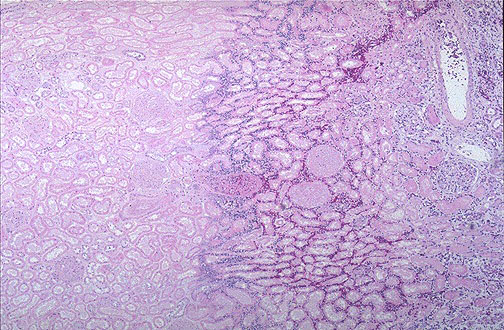
16.Microscopically, the renal cortex has undergone anoxic injury at the left so that the cells appear pale and ghost-like. There is a hemorrhagic zone in the middle where the cells are dying or have not quite died, and then normal renal parenchyma at the far right. This is an example of coagulative necrosis.
16.镜下,左边肾皮质缺氧坏死,细胞染色变淡。中间有出血区,细胞坏死不彻底。最右边是正常的肾皮质。本图为凝固性坏死。